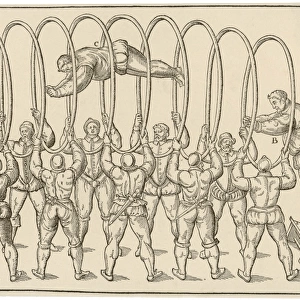
Home > Arts > Artists > J > Jacob Jacobs
Dental anatomy

![]()
Wall Art and Photo Gifts from Science Photo Library
Dental anatomy
Dental anatomy. Historical anatomical artwork of healthy and diseased human teeth and jaws. The teeth and jaws are seen from several different directions. At upper right is an adults upper jaw, seen from below. At lower right is an adults lower jaw, seen from above. At centre left are an adults jaws seen from the side, dissected to show the roots of the teeth. At lower left are two views of a childs jaws showing the milk teeth and buds of adult teeth. The artworks of individual teeth are mostly diseased or deformed, like the molars at upper centre with fused or divergent roots. Artwork from Atlas of Anatomy, by Bourgery and Jacob, published in France in 8 volumes from 1831 to 1854
Science Photo Library features Science and Medical images including photos and illustrations
Media ID 6447971
© MEHAU KULYK/SCIENCE PHOTO LIBRARY
Anatomical Artwork Child Conditions Dental Dentistry Diseased Diseases Disorders Dissected Dissection Drawing Jacob Jaws Jean Baptiste Marc Bourgery Mandible Mouth Nicolas Henri Jacob Oral Teeth Tooth
FEATURES IN THESE COLLECTIONS
> Arts
> Artists
> J
> Jacob Jacobs
EDITORS COMMENTS
This print showcases a remarkable piece of historical anatomical artwork, highlighting the intricate details of dental anatomy. The image captures both healthy and diseased human teeth and jaws from various perspectives, providing a comprehensive view of this vital aspect of our bodies. At the upper right corner, we are presented with an adults' upper jaw seen from below, while at the lower right corner, an adults' lower jaw is depicted from above. In the center left section, we can observe an adult's dissected jaws revealing the roots of the teeth. Lastly, in the lower left portion, two views of a child's jaws display both milk teeth and budding adult teeth. The artwork itself predominantly focuses on diseased or deformed individual teeth. Notably, at the upper center lies a depiction of molars with fused or divergent roots—a fascinating representation that highlights dental conditions prevalent during that era. Originally published in France between 1831 to 1854 as part of Jean Baptiste Marc Bourgery and Nicolas Henri Jacob's Atlas of Anatomy—an eight-volume masterpiece—this illustration offers valuable insights into dentistry's historical development. With its blend of artistry and scientific accuracy, this print serves as a testament to how our understanding and treatment methods for dental diseases have evolved over time. It stands as a visual reminder not only of our oral health but also our continuous pursuit to unravel the mysteries hidden within our own bodies.
MADE IN THE USA
Safe Shipping with 30 Day Money Back Guarantee
FREE PERSONALISATION*
We are proud to offer a range of customisation features including Personalised Captions, Color Filters and Picture Zoom Tools
SECURE PAYMENTS
We happily accept a wide range of payment options so you can pay for the things you need in the way that is most convenient for you
* Options may vary by product and licensing agreement. Zoomed Pictures can be adjusted in the Cart.