Home > Science > SEM
Trachea lining, SEM C015 / 9936

![]()
Wall Art and Photo Gifts from Science Photo Library
Trachea lining, SEM C015 / 9936
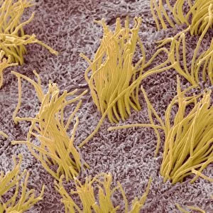
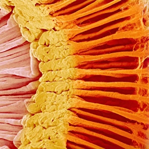
Trachea lining. Coloured scanning electron micrograph (SEM) of a section through the lining of the trachea (wind pipe). The trachea links the larynx (voice box) to the lungs. Across upper frame are pseudostratified ciliated columnar epithelial cells (bright orange), which line the trachea. The hair-like cilia (yellow) on their surface beat to move mucous, and particles trapped in it, upwards out of the respiratory tract. This helps to keep the lungs and airways clear and prevent infection. At bottom is the basement membrane, a layer of connective tissue that supports the epithelial cells
Science Photo Library features Science and Medical images including photos and illustrations
Media ID 9239439
© SUSUMU NISHINAGA/SCIENCE PHOTO LIBRARY
Basement Membrane Cilia Columnar Epithelium Connective Tissue Epithelial Epithelium Lining Respiratory Scanning Microscopy Trachea Tract Wind Pipe Cells Section Sectioned
EDITORS COMMENTS
This print showcases the intricate beauty of the trachea lining, as seen through a colored scanning electron microscope (SEM). The trachea, commonly known as the windpipe, serves as a vital connection between the larynx and lungs. In this image, we observe the upper frame adorned with bright orange pseudostratified ciliated columnar epithelial cells that form a protective layer along the tracheal lining. The mesmerizing yellow hair-like structures on their surface are none other than cilia. These tiny projections tirelessly beat in unison to propel mucus and any trapped particles upwards and out of our respiratory tract. This remarkable mechanism plays a crucial role in maintaining clear airways and preventing infections within our lungs. At the bottom of this visual masterpiece lies the basement membrane—a supportive layer composed of connective tissue that provides stability to these delicate epithelial cells. Together, they create an extraordinary harmony necessary for optimal respiratory function. Through SUSUMU NISHINAGA's lens and expertise in scanning electron microscopy, we gain an awe-inspiring glimpse into this microscopic world within us. This photograph not only highlights the intricacies of biology but also reminds us of our body's remarkable ability to protect itself from harm.
MADE IN THE USA
Safe Shipping with 30 Day Money Back Guarantee
FREE PERSONALISATION*
We are proud to offer a range of customisation features including Personalised Captions, Color Filters and Picture Zoom Tools
SECURE PAYMENTS
We happily accept a wide range of payment options so you can pay for the things you need in the way that is most convenient for you
* Options may vary by product and licensing agreement. Zoomed Pictures can be adjusted in the Cart.